THE VALUE OF BRAIN MRI IN SCREENING AND EARLY DETECTION OF GENETICALLY PREDISPOSED CEREBRAL ANEURYSMS
5/18/2026 9:58:39 AM
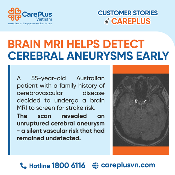
A 55-year-old female patient from Australia, while traveling in Vietnam, visited CarePlus to undergo a brain Magnetic Resonance Imaging (MRI) scan. During the pre-scan consultation with the radiologist, the patient explained that her decision to undergo screening stemmed from a significant family event: her older sister had suddenly passed away from a ruptured cerebral aneurysm the previous year. Given this family history and genetic risk factor, the patient sought an in-depth evaluation to proactively manage her health risks.
IMAGING FINDINGS (MRI)
MRI assessment using the 3D Time-of-Flight Magnetic Resonance Angiography (3D-TOF MRA) sequence (non-contrast cerebral vascular imaging) revealed an unruptured intracranial aneurysm:
- Location: Right internal carotid artery aneurysm, supraclinoid segment.
- Morphology: Saccular aneurysm with a wide neck (wide-necked aneurysm), measuring approximately 5.5 mm in maximum diameter.
- Brain parenchyma: No evidence of hemorrhage, mass effect, or compression of adjacent neural structures was identified.
PROFESSIONAL ANALYSIS
The use of MRI in this case provided significant preventive diagnostic value, enabling the detection of silent vascular abnormalities before catastrophic complications occurred.
1. Non-invasive vascular assessment – The advantage of 3D-TOF MRA
Unlike CT Angiography (CTA) or Digital Subtraction Angiography (DSA), 3D-TOF MRA utilizes the natural flow of blood to reconstruct vascular anatomy.
Clinical significance: This is considered one of the safest screening modalities for asymptomatic patients. It allows detailed evaluation of the cerebral vascular system without ionizing radiation or contrast injection, making it especially suitable for periodic screening in individuals with a family history of cerebral aneurysms.
2. Characterization of aneurysm features and risk stratification
MRI enabled precise assessment of the aneurysm’s size (5.5 mm), location (supraclinoid segment), and morphology (wide-neck configuration).
Clinical implications: From a medical perspective, not all cerebral aneurysms require immediate intervention. Many small aneurysms may remain stable throughout life. However, the combination of a 5.5 mm aneurysm, wide-neck morphology, and positive first-degree family history represents important risk indicators that guide specialists toward close surveillance or preventive intervention before rupture occurs.
3. Early detection – The key to reducing mortality risk
MRI’s high sensitivity in detecting unruptured aneurysms offers patients a valuable “window of opportunity.” Early diagnosis removes uncertainty regarding health status and shifts management from a reactive approach to proactive risk control.
CLINICAL VALUE OF MRI
In diagnostic imaging, MRI not only detects abnormalities but also provides a detailed “pathological roadmap” for subsequent clinical management. For this patient, the findings identified in Vietnam did not constitute an immediate emergency; rather, they represented invaluable medical information.
With these imaging results, the patient can return to Australia well-prepared for consultation with neurosurgeons or neurovascular intervention specialists. Based on the clearly documented imaging findings, an individualized management plan — ranging from periodic monitoring to advanced endovascular interventions such as stent placement or coil embolization — can be established.
Close monitoring and timely intervention based on early screening are essential in preventing subarachnoid hemorrhage, reducing mortality risk, and preserving the patient’s long-term quality of life.