Pelvic MRI - Contrast Enhancement
1. What is Pelvic MRI (Prostate, Uterus, Ovaries Examination)?
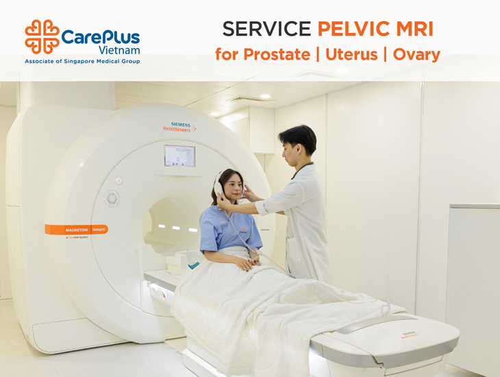
Pelvic MRI is an advanced imaging technique that uses strong magnetic fields and radio waves to produce detailed images of pelvic structures, including the prostate in men and the uterus and ovaries in women. This method does not involve X-rays, ensuring safety, and provides high-resolution images to help doctors detect lesions, tumors, or abnormalities in the pelvic region.
2. When is Pelvic MRI Necessary?
Pelvic MRI is commonly prescribed in the following cases:
Prostate Disorders: To evaluate prostate cancer, prostatitis, or benign prostatic hyperplasia (BPH).
Gynecological Screening: To identify conditions such as uterine fibroids, endometriosis, ovarian cysts, or uterine/ovarian cancer.
Pelvic Pain Assessment: To investigate causes of chronic or acute pelvic pain when other diagnostic methods are inconclusive.
Treatment Planning: To aid in planning surgeries or radiation therapy for cancers or other pelvic conditions.
Post-Treatment Monitoring: To evaluate the effectiveness of surgery, chemotherapy, or radiation therapy.
3. Who Should Undergo Pelvic MRI?
Men: Those at high risk for or diagnosed with prostate conditions.
Women: Individuals with menstrual irregularities, unexplained lower abdominal pain, or suspected ovarian/uterine diseases.
Cancer Patients: Those with a history of pelvic cancer requiring recurrence monitoring.
Patients with Symptoms: Experiencing difficulty urinating, pelvic pain, or unexplained blood in urine.
4. Advantages and Precautions of Pelvic MRI
Advantages:
- Radiation-Free: Safe for health as it does not use X-rays.
- High-Resolution Images: Provides superior diagnostic accuracy compared to ultrasound or CT scans.
- Early Detection: Capable of identifying small abnormalities or early-stage diseases.
Precautions:
- Inform Your Doctor: Disclose if you are pregnant, have implanted metal devices, or suffer from kidney conditions (if contrast dye is used).
- Follow Instructions: Adhere to guidelines such as fasting before the procedure if instructed.
- Stay Still During the Scan: Minimize movement to ensure clear and accurate imaging.
5. Conclusion
Pelvic MRI is a safe and effective diagnostic tool, offering detailed insights into pelvic health. It facilitates early detection, accurate diagnosis, and informed treatment planning for critical conditions, contributing to improved health outcomes and quality of life.