LATERAL KNEE PAIN AFTER A TWISTING INJURY – A “HIDDEN CORNER” INJURY THAT IS EASILY MISSED
2/26/2026 9:59:01 AM
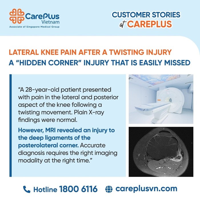
Ms. H.T.K.A., a 28-year-old office worker, sustained a knee injury during sports activity. While performing a sudden twisting movement with her lower leg fixed on the ground, she heard a faint popping sound on the lateral aspect of her left knee. Immediately afterward, she experienced sharp pain along the lateral and posterior aspects of the knee.
Over the following days, the pain was no longer severe but persisted as a dull ache, worsening particularly when squatting or rotating the leg. Initial plain X-ray examination was completely normal, with no evidence of fracture. Suspecting a ligamentous injury, the orthopedic specialist ordered an MRI of the knee joint.
MRI FINDINGS OF THE KNEE
- MRI accurately identified a deep-seated injury in the posterolateral corner (PLC) — an anatomically complex and often overlooked region:
- Primary ligament injury: Partial tear of the popliteofibular ligament, accompanied by surrounding soft-tissue edema.
- Bone involvement: Mild bone marrow edema at the proximal fibular head, corresponding to the ligament and tendon attachment sites.
- Joint reaction: Small amount of fluid in the suprapatellar bursa and minimal knee joint effusion.
THE VALUE OF MRI IN KNEE JOINT PATHOLOGY
In this case, MRI played a decisive “referee” role:
- Detection of subtle injuries in difficult locations: The popliteofibular ligament is small and deeply located, making it extremely difficult to assess accurately with ultrasound or X-ray. MRI is the only modality capable of clearly demonstrating partial tears and soft-tissue edema in the posterolateral corner.
- Assessment of bone marrow edema: MRI confirmed bone injury caused by traction or impact forces, despite the absence of fracture on plain radiographs.
- Guidance for treatment planning: Identifying the injury as a partial tear (rather than a complete rupture) allowed clinicians to confidently choose conservative management—such as knee bracing and avoidance of rotational movements—enabling ligament healing and reducing the risk of future chronic knee instability.
EXPERT ADVICE
Although MRI has significant value in detecting deep ligamentous injuries like in Ms. A.’s case, not every patient with knee pain requires immediate MRI.
Routine overuse of MRI can lead to unnecessary healthcare costs. In many common cases of knee pain due to superficial soft-tissue trauma, a thorough clinical examination combined with ultrasound is often sufficient for diagnosis.
Conclusion
The choice of imaging modality—whether X-ray, ultrasound, or MRI—should be determined by a musculoskeletal specialist. Close collaboration between the clinical physician and the radiologist is the key factor in establishing the most accurate treatment plan and avoiding missed subtle injuries in the posterolateral corner of the knee.