THE VALUE OF MRI IN COMPREHENSIVE ASSESSMENT OF POST-TRAUMATIC SHOULDER INSTABILITY
7/1/2026 9:11:17 AM
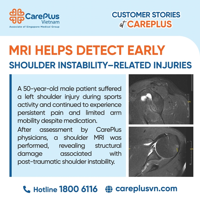
A 50-year-old male patient presented with a history of direct trauma to the left shoulder after falling onto an outstretched arm while playing sports. Following the injury, he developed progressively worsening dull shoulder pain. Despite three weeks of self-treatment with pain medication and rest, his symptoms failed to improve and became more severe, with significant restriction of shoulder range of motion, particularly during abduction and arm elevation. Given the clinical suspicion of a deep structural injury involving the shoulder complex, the physician recommended Magnetic Resonance Imaging (MRI) for detailed evaluation.
MRI FINDINGS
MRI of the left shoulder revealed a combination of complex osseous and soft-tissue injuries, characteristic of post-dislocation (or subluxation) shoulder instability:
- Bony Bankart lesion: Disruption of the anteroinferior glenoid rim with an associated bony fragment, accompanied by injury to the corresponding labral structure.
- Hill-Sachs lesion: A mild compression defect was identified at the posterolateral aspect of the humeral head.
- Inferior Glenohumeral Ligament (IGHL) injury: Complete tear of the IGHL complex, the primary stabilizing structure that maintains shoulder stability when the arm is abducted and externally rotated.
- Inflammatory response: Moderate fluid accumulation within the subcoracoid bursa extending along the bicipital groove, accompanied by bone marrow edema at sites of osseous impact.
PROFESSIONAL ANALYSIS
In this case, MRI provided decisive diagnostic value by revealing injuries that are often overlooked or incompletely assessed by conventional radiography or ultrasound.
1. Multilevel Assessment of Injury: The “Bankart–Hill-Sachs” Combination
MRI confirmed the presence of both a bony Bankart lesion and a Hill-Sachs lesion. Together, these findings serve as highly specific indirect evidence of a previous anterior shoulder dislocation occurring at the time of injury.
Clinical significance:
When the humeral head dislocates anteriorly from the glenoid cavity, its posterolateral surface impacts against the anterior glenoid rim, creating the characteristic Hill-Sachs defect and potentially fracturing the glenoid rim. A bony Bankart lesion reduces the articular surface area of the glenoid, predisposing the patient to chronic shoulder instability if not properly treated.
MRI enables accurate quantification of bone loss and defect size, providing essential information for orthopedic surgeons when determining whether labral repair alone is sufficient or whether bone-grafting procedures are required.
2. Evaluation of the Shoulder’s Key Stabilizer: The IGHL
Detection of an Inferior Glenohumeral Ligament (IGHL) tear represents one of MRI’s most valuable contributions in shoulder instability assessment. The IGHL functions like a supportive “hammock” beneath the shoulder joint and serves as the most important ligamentous restraint against anterior and inferior subluxation or dislocation.
Clinical implications: Loss of this critical stabilizing structure explains the patient’s severe pain and functional impairment. If an IGHL rupture is overlooked, the patient is at high risk of developing chronic instability and recurrent shoulder dislocations.
3. Detection of Bone Marrow Edema and Diffuse Bursitis
MRI’s high sensitivity to water content allows visualization of bone marrow edema, indicating ongoing mechanical injury and tissue response following trauma.
The extensive fluid collection extending from the subcoracoid bursa into the bicipital groove accounts for the patient's persistent and significant pain. This inflammatory reaction is secondary to trauma and may contribute to the development of secondary adhesive capsulitis (“frozen shoulder”) if timely rehabilitation and physical therapy are not initiated.
THE CLINICAL VALUE OF MRI
In shoulder trauma assessment, while radiographs are useful for excluding major fractures, MRI is widely regarded as the gold standard for evaluating soft-tissue structures, ligaments, cartilage, and labral injuries.
For this patient, the persistence of symptoms beyond three weeks was not simply attributable to a routine soft-tissue injury, but rather reflected significant structural instability of the shoulder joint.
MRI not only established the diagnosis but also provided a comprehensive “anatomical roadmap” of the injury, detailing osseous lesions (Hill-Sachs and Bankart injuries), ligamentous damage (IGHL rupture), and associated inflammatory changes including bone marrow edema.
Early intervention guided by MRI findings—ranging from appropriate immobilization to arthroscopic reconstructive surgery—is critical for preserving shoulder function, restoring joint stability, and preventing long-term complications such as recurrent dislocation and premature shoulder osteoarthritis.